Abdominal Anatomy - Abdominal Anatomy Gif Png Download Illustration Clipart 5141625 Pikpng / The anterolateral abdominal wallformed of 4 layer skin, fascia, muscles, and peritoneum.. The anterolateral abdominal wall consists of four main layers (external to internal): The major organs of the abdomen include the small intestine, large intestine, and stomach. If you plan to enter a healthcare profession such as nursing, this is something you'll use on the job when performing abdominal assessments (and while documenting). It extends to the lumbar spine, which joins the thorax and pelvis and is a point of attachment for some abdominal wall structures 1 . Learn the anatomy and function of your abdominals to achieve your dream physique.
The area occupied by the abdomen is called the abdominal cavity. The majority of these organs are encased in a protective membrane termed the peritoneum. If you plan to enter a healthcare profession such as nursing, this is something you'll use on the job when performing abdominal assessments (and while documenting). The anterolateral abdominal wallformed of 4 layer skin, fascia, muscles, and peritoneum. This course covers all essentials:
Abdominal Surface Anatomy Creative Commons Illustration Radiology Case Radiopaedia Org from prod-images-static.radiopaedia.org The abdomen contains many vital organs: For the sake of brevity, the various organs will be not discussed in detail. The abdomen is the body region found between the thorax and the pelvis. Browse 7,542 abdomen anatomy stock photos and images available, or search for digestive system to find more great stock photos and pictures. It is bounded superiorly by the xiphoid process and costal margins, posteriorly by the vertebral column and inferiorly by the pelvic bones and inguinal ligament. Learn the anatomy and function of your abdominals to achieve your dream physique. The majority of these organs are encased in a protective membrane termed the peritoneum. Skin, superficial fascia, muscles and associated fascia, and parietal peritoneum.
Its superior aperture faces towards the thorax, enclosed by the diaphragm.
Stomach is a muscular bag forming the most distensible part of the human digestive system. Outline • abdominal cavity • external and internal fascia • abdominal muscle • intraperitoneal and retroperitoneal organs • ligaments • topography • laparotomy and its sites • summary It is bounded superiorly by the xiphoid process and costal margins, posteriorly by the vertebral column and inferiorly by the pelvic bones and inguinal ligament. For the sake of brevity, the various organs will be not discussed in detail. The component of the urinary system, kidney and the ureter. The major organs of the abdomen include the small intestine, large intestine, and stomach. Together, these three turn nutrients into usable energy, as well as help dispose of solid waste. It extends to the lumbar spine, which joins the thorax and pelvis and is a point of attachment for some abdominal wall structures 1 . The posterior abdominal wall is a musculoskeletal structure formed by the posterior abdominal muscles, their fascia, the lumbar vertebrae and the pelvic girdle. The stomach, the small intestine (jejunum and ileum), the large intestine (colon), the liver, the spleen, the gallbladder, the pancreas, the uterus, the fallopian tubes, the ovaries, the kidneys, the ureters, the bladder, and many blood vessels (arteries and veins). The abdomen is the front part of the abdominal segment of the trunk. The area occupied by the abdomen is called the abdominal cavity. This course covers all essentials:
The anterolateral abdominal wall consists of four main layers (external to internal): The component of the urinary system, kidney and the ureter. The major organs of the abdomen include the small intestine, large intestine, and stomach. Together, these three turn nutrients into usable energy, as well as help dispose of solid waste. Skin, superficial fascia, muscles and associated fascia, and parietal peritoneum.
Right Upper Quadrant Anatomy And Causes For Pain Kenhub from thumbor.kenhub.com The muscle fibers of the transversus abdominis run horizontally, similar to a corset or a weight belt. You can't have a strong, muscular physique without a healthy, stable core. The stomach, the small intestine (jejunum and ileum), the large intestine (colon), the liver, the spleen, the gallbladder, the pancreas, the uterus, the fallopian tubes, the ovaries, the kidneys, the ureters, the bladder, and many blood vessels (arteries and veins). In anatomy and physiology, you'll learn how to divide the abdomen into nine different regions and four different quadrants. The abdominal aorta enters the abdomen through the diaphragm at the level of the twelfth thoracic vertebre and continues to just below the umbilical area, where it splits into the right and left common iliac arteries. The abdomen contains many vital organs: Phil anatomy (3rd ) 2. Abdominal computed tomography (ct) is a type of medical imaging procedure used to diagnose and monitor internal stomach issues, like cancer, bowel obstruction, and abdominal pain.
Inferiorly the abdomen is open to the pelvis, communicating through the superior pelvic aperture (pelvic inlet).
The region occupied by the abdomen is called the abdominal cavity, and is enclosed by the abdominal muscles at front and to the sides, and by part of the vertebral column at the back. The abdomen (colloquially called the belly, tummy, midriff or stomach) is the part of the body between the thorax (chest) and pelvis, in humans and in other vertebrates. The component of the urinary system, kidney and the ureter. The aorta is the largest blood vessel in the body. Phil anatomy (3rd ) 2. It also contains the suprarenal glands and the major neurovascular. Abdomen anatomy the abdomen is comprised primarily of the digestive tract and other accessory organs which assist in digestion, the urinary system, spleen, and the abdominal muscles (shown below). This muscle doesn't help move the spine or the pelvis, but it does help with respiration and breathing. The anterolateral abdominal wallformed of 4 layer skin, fascia, muscles, and peritoneum. If you plan to enter a healthcare profession such as nursing, this is something you'll use on the job when performing abdominal assessments (and while documenting). The anterolateral abdominal wall consists of four main layers (external to internal): We'll identify as many organs as we can, see how they fit into the. It extends to the lumbar spine, which joins the thorax and pelvis and is a point of attachment for some abdominal wall structures 1 .
Phil anatomy (3rd ) 2. Its superior aperture faces towards the thorax, enclosed by the diaphragm. This course covers all essentials: The regions occupied by stomach are epigastric, umbilical and hypochondriac regions. The major organs of the abdomen include the small intestine, large intestine, and stomach.
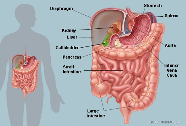
The Abdomen Human Anatomy Picture Function Parts Definition And More from img.webmd.com The abdominal aorta enters the abdomen through the diaphragm at the level of the twelfth thoracic vertebre and continues to just below the umbilical area, where it splits into the right and left common iliac arteries. It extends to the lumbar spine, which joins the thorax and pelvis and is a point of attachment for some abdominal wall structures 1 . These organs are held together loosely by connecting tissues. Abdomen, in human anatomy, the body cavity lying between the chest or thorax above and the pelvis below and from the spine in the back to the wall of abdominal muscles in the front. The majority of these organs are encased in a protective membrane termed the peritoneum. The abdomen is the part of the body that contains all of the structures between the thorax (chest) and the pelvis, and is separated from the thorax via the diaphragm. The component of the urinary system, kidney and the ureter. It also contains the spleen.
Anatomy of the posterior abdominal wall the abdominal cavity is bounded by the abdominal wall which is divided into an anterior wall, lateral wall and the posterior wall.
Anatomy of the posterior abdominal wall the abdominal cavity is bounded by the abdominal wall which is divided into an anterior wall, lateral wall and the posterior wall. The major organs of the abdomen include the small intestine, large intestine, and stomach. It also contains the spleen. In anatomy and physiology, you'll learn how to divide the abdomen into nine different regions and four different quadrants. The abdominal region is supported by the anterior and posterior abdominal wall that supports the viscera and maintains the posturewhere there's no bony support. The muscle fibers of the transversus abdominis run horizontally, similar to a corset or a weight belt. The aorta is the largest blood vessel in the body. The component of the urinary system, kidney and the ureter. The area occupied by the abdomen is called the abdominal cavity. The transverse abdominal muscle wraps around the torso from front to back and from the ribs to the pelvis. The abdominal wall surrounds the abdominal cavity, providing it with flexible coverage and protecting the internal organs from damage. It is an artery, meaning that it carries blood away from the heart. Browse 7,542 abdomen anatomy stock photos and images available, or search for digestive system to find more great stock photos and pictures.